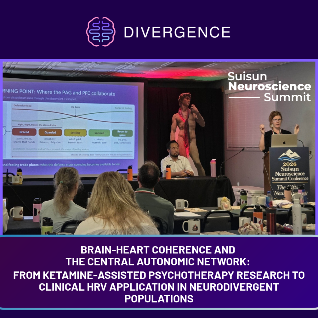
Brain-heart coherence describes the relationship between neural activity and autonomic regulation. The central autonomic network (CAN), a distributed system involving the prefrontal cortex, anterior cingulate cortex, insula, amygdala, hypothalamus, and brainstem, contributes to the integration of emotional, cognitive, and autonomic processes (Lamotte et al., 2021). EEG and heart rate variability (HRV) provide complementary perspectives on these processes. EEG measures electrical brain activity, while HRV measures variation in the intervals between heartbeats. Examining both signals can help researchers and clinicians understand how neural and autonomic activity relate across different physiological and clinical states.
At the 2026 Suisun Neuroscience Summit: The Gifts of Neurodiversity, Heather Hargraves, M.A., C.Psych., and Alex Ni, MBA, CPA, explored this relationship in their presentation, “Brain-Heart Coherence and the Central Autonomic Network: From Ketamine-Assisted Psychotherapy Research to Clinical HRV Application in Neurodivergent Populations.” The presentation connected observational findings from ketamine-assisted psychotherapy (KAP) with the clinical application of HRV in neurodivergent and trauma-related presentations, framing brain-heart coherence as a dynamic relationship influenced by physiological state, context, and individual differences.
What Is the Central Autonomic Network?
The central autonomic network integrates information related to emotion, cognition, internal physiological state, and autonomic function. Structures including the insula, anterior cingulate cortex, amygdala, hypothalamus, and brainstem contribute to this system (Lamotte et al., 2021). The neurovisceral integration model similarly describes interactions between prefrontal, limbic, and autonomic systems involved in emotional regulation (Thayer & Lane, 2000).
HRV provides a peripheral measure that can offer information about these processes. It reflects variation in the time between heartbeats and is influenced by respiration, posture, activity, medication, age, and health status (Shaffer & Ginsberg, 2017). HRV should therefore not be treated as a direct measurement of the CAN or as a standalone measure of regulation. Its clinical value depends on interpreting changes within the broader physiological and clinical context.
What Ketamine-Assisted Psychotherapy Reveals About Neural Complexity
The KAP research presented by Hargraves and Ni drew on an observational study of healthy volunteers undergoing ketamine-assisted psychotherapy in Frankfurt, Germany, in October 2025, conducted in collaboration with Dr. Mario Scheib. Simultaneous EEG and HRV monitoring was used to examine neural and autonomic activity during the ketamine-assisted state, with Lempel-Ziv Complexity (LZC) used to assess neural signal complexity.
The observations showed increased sympathetic activation alongside increased neural complexity. This combination raised questions about models in which greater HRV and neural flexibility are expected to move together, suggesting that relationships between neural complexity and autonomic activity may change during altered physiological states.
These observations were preliminary and did not establish a general mechanism for how ketamine affects brain-heart communication. Their value lay in examining how neural and autonomic dynamics may interact during KAP and whether these relationships differ from those observed during ordinary waking states.
How EEG and HRV May Relate Across Frequency Bands
The presentation also examined relationships between cardiac frequency components and EEG activity. High-frequency HRV activity showed closer associations with changes in frontal alpha and theta complexity, while low-frequency dynamics demonstrated different associations with beta-range cortical activity.
These findings suggested that brain-heart relationships may vary across frequency ranges rather than functioning as a single uniform interaction. The findings were best considered exploratory evidence of associations between cardiac and cortical activity.
HRV and Physiological Context in Neurodivergent Populations
HRV can provide physiological information that complements subjective reporting and behavioral observation. Research has examined autonomic activity across autism and ADHD, but findings vary according to the population, HRV measure, task, environment, and other physiological and methodological factors.
For clinicians, this makes HRV more useful as part of a broader assessment than as a diagnostic marker. Cardiac variability can be considered alongside EEG measures, observed behavior, subjective experience, and the demands placed on the individual. This shifts the question from whether HRV is simply high or low to how physiological activity changes under different conditions.
Trauma, Dissociation, and Physiological Regulation
Trauma-related presentations further demonstrate the importance of clinical context. Thome et al. (2022) examined associations between HRV and brainstem-limbic connectivity in PTSD and its dissociative subtype and found different patterns of association across groups. Although exploratory, the study illustrates how similar clinical presentations may involve different relationships between autonomic and neural systems.
This is particularly relevant when considering states such as hyperarousal and dissociation. HRV cannot independently determine which state a person is experiencing, but physiological measurements may provide additional information when interpreted alongside clinical assessment and subjective experience.
Alexithymia, Interoception, and Subjective Experience
The relationship between physiological signals and subjective experience is also relevant to alexithymia and interoception. Alexithymia involves difficulty identifying and describing emotions, while interoception encompasses several distinct abilities. Garfinkel et al. (2015) distinguish between interoceptive accuracy, interoceptive sensibility, and interoceptive awareness, demonstrating why physiological measurements and perceived internal states should not automatically be assumed to correspond.
This distinction is relevant to biofeedback. Physiological information can provide another reference point for exploring changes in arousal and emotion without treating the physiological signal as inherently more accurate than the client’s report. Differences between physiological and subjective measures may themselves provide useful information within the therapeutic process.
Developing Individualized HRV and Neurofeedback Training
These concepts can inform an integrated approach to neurofeedback and biofeedback for ADHD, autism, and trauma-related presentations. Rather than applying the same training strategy across populations, the emphasis is on sequencing and individualization. Baseline physiology, responses to different demands, subjective experience, EEG patterns, and changes across sessions can all contribute to decisions about how training is introduced and adjusted.
This approach also avoids treating higher HRV or greater neural complexity as automatic indicators of better functioning. A more useful clinical goal is to understand how an individual’s neural and autonomic systems respond to changing demands and whether training is associated with greater adaptability within that person’s context.
Bringing EEG and HRV Together in Clinical Practice
EEG and HRV provide complementary information. EEG can be used to examine cortical activity and measures such as neural complexity, while HRV provides information about cardiac variability and autonomic function. Subjective reporting adds another perspective on how the individual experiences these changes.
Using these measures together does not mean that one signal directly explains another. Instead, their combination can help clinicians examine how neural activity, autonomic responses, and subjective experience change across conditions and training sessions. This can support a more individualized understanding of treatment response without relying on a single physiological metric.
What Brain-Heart Coherence Means for Clinical Practice
Brain-heart coherence is best understood as a dynamic relationship rather than a single number or ideal physiological state. The work presented by Heather Hargraves and Alex Ni brought together research on the CAN, neural complexity, HRV, altered states, neurodivergence, trauma, and interoception to examine how neural and autonomic systems may interact under different conditions.
The clinical question is therefore not simply whether the brain and heart are “in sync.” It is how neural and autonomic activity relate as an individual responds to changing demands, and whether those relationships can provide useful information for personalized neurofeedback and biofeedback training.
Frequently Asked Questions
Q: What is brain-heart coherence?
A: Brain-heart coherence refers to the relationship between neural activity and cardiovascular or autonomic activity. It is a framework for studying brain-body regulation rather than a single physiological measurement.
Q: What is the Central Autonomic Network?
A: The Central Autonomic Network is a distributed network of brain regions involved in autonomic and emotional regulation, including the insula, anterior cingulate cortex, amygdala, hypothalamus, and brainstem structures (Lamotte et al., 2021).
Q: What does HRV measure?
A: Heart rate variability measures variation in the intervals between consecutive heartbeats. It provides information about cardiac and autonomic function but is influenced by multiple physiological and environmental factors (Shaffer & Ginsberg, 2017).
Q: Can HRV be used with neurodivergent populations?
A: HRV can provide physiological information when working with neurodivergent populations, but research findings vary across autism and ADHD. It should not be used as a standalone diagnostic measure and is better interpreted alongside clinical, behavioral, and subjective information.
Q: Why combine EEG and HRV?
A: EEG and HRV measure different physiological processes. Using them together allows clinicians and researchers to examine relationships between cortical activity and cardiovascular variability that may not be apparent when either signal is considered independently.
Stay Informed on the Future of Neurofeedback and Biofeedback
The Divergence Neuro newsletter, NeuroNotes, delivers evidence-informed insights on nervous system regulation, trauma-informed care, emerging technologies, and practical applications in neurofeedback and biofeedback.
Subscribe to NeuroNotes for the latest research, clinical insights, and developments in the field.
Physiological measures such as EEG and HRV are intended to support, not replace, professional clinical assessment and judgment.
References
Alba, G., Vila, J., Rey, B., Montoya, P., & Muñoz, M. Á. (2019). The relationship between heart rate variability and electroencephalography functional connectivity variability is associated with cognitive flexibility. Frontiers in Human Neuroscience, 13, 64. https://doi.org/10.3389/fnhum.2019.00064
Chang, C., Metzger, C. D., Glover, G. H., Duyn, J. H., Heinze, H.-J., & Walter, M. (2013). Association between heart rate variability and fluctuations in resting-state functional connectivity. NeuroImage, 68, 93–104. https://doi.org/10.1016/j.neuroimage.2012.11.038
Garfinkel, S. N., Seth, A. K., Barrett, A. B., Suzuki, K., & Critchley, H. D. (2015). Knowing your own heart: Distinguishing interoceptive accuracy from interoceptive awareness. Biological Psychology, 104, 65–74. https://doi.org/10.1016/j.biopsycho.2014.11.004
Lamotte, G., Shouman, K., & Benarroch, E. E. (2021). Stress and central autonomic network. Autonomic Neuroscience: Basic and Clinical, 235, 102870. https://pubmed.ncbi.nlm.nih.gov/34461325/
Medel, V., Irani, M., Crossley, N., Ossandón, T., & Boncompte, G. (2023). Complexity and 1/f slope jointly reflect brain states. Scientific Reports, 13, 21700. https://doi.org/10.1038/s41598-023-47316-0
Shaffer, F., & Ginsberg, J. P. (2017). An overview of heart rate variability metrics and norms. Frontiers in Public Health, 5, 258. https://doi.org/10.3389/fpubh.2017.00258
Thayer, J. F., & Lane, R. D. (2000). A model of neurovisceral integration in emotion regulation and dysregulation. Journal of Affective Disorders, 61(3), 201–216. https://pubmed.ncbi.nlm.nih.gov/11163422/
Thome, J., Densmore, M., Terpou, B. A., Théberge, J., McKinnon, M. C., & Lanius, R. A. (2022). Contrasting associations between heart rate variability and brainstem-limbic connectivity in posttraumatic stress disorder and its dissociative subtype: A pilot study. Frontiers in Behavioral Neuroscience, 16, 862192. https://doi.org/10.3389/fnbeh.2022.862192